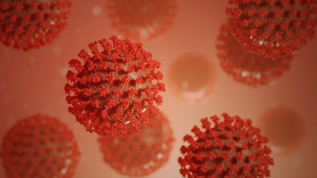
Coronaviruses(COVID-19) are a group of related RNA viruses that cause diseases in mammals and birds. In humans, these viruses are cause respiratory tract infections that can be range from mild to lethal. Mild illnesses include some common cold cases, while more lethal varieties can cause SARS, MERS, and COVID-19.
Symptoms in other species vary: in chicken, they produce an upper respiratory tract disease, while in cows and pigs, they cause diarrhea. There are as yet no vaccines and antiviral drugs to prevent or treat human coronavirus(COVID-19) infections.
Coronavirus constitutes of the subfamily Orthocoronavirinae, in the family Coronaviridae, order Nidovirales, and realm Riboviria. They have also enveloped viruses with a positive-sense single-stranded RNA genome and a nucleocapsid of helical symmetry.
This is wrapped in an icosahedral protein shell. The genome size of the coronaviruses ranges from approximately 26 to 32 kilobase, one of the largest among RNA viruses. They have characteristic club-shaped spikes that project from their surfaces, which is an electron micrograph that create an image reminiscent of the solar corona, from which their name derives.
Etymology
The name of coronavirus(COVID-19) is derived from Latin corona, meaning "crown" or "wreath," itself a borrowing from GreekS korṓnē, "garland, wreath."The name was coined by June Almeida and David Tyrrell, who first observed and studied human coronaviruses.
The word was the first used in print in 1968 by an informal group of virologists in the journal Nature of designate the new family of viruses.
The name refers to the characteristic appearance of virions by electron microscopy, which has a fringe of large, bulbous surface projections creating an image reminiscent of the solar corona or halo. This morphology is created by the viral spike peplomer, which are proteins on the surface of the viruses.
History
Coronaviruses were first discovered in the 1930s when an acute respiratory infection of a domesticated chicken was caused by the infectious bronchitis virus (IBV). Arthur Schalk and M.C. Hawn described in 1931 new respiratory infections of chickens in North Dakota.
The diseases of new-born chicks were characterized by gasping and listlessness. The chicks' mortality rate was 40–90%. Six years later, Fred Beaudete and Charles Hudson successfully isolated and cultivated the infectious bronchitis virus, which caused the disease.
In the 1940s, two more on animal coronaviruses, mouse hepatitis virus, and transmissible gastroenteritis virus, were isolated. It was not realized at the time that these three differents viruses were related.Human coronaviruses were discovered within the Sixties.
They were isolated victimisation 2 totally different ways within the uk and also the u. s.. E.C. Malcom Byone and Davids Tyrrell are working at the Common Colds Unit of the British Medical Research Council since 1960, isolated from a boy a novel common colds virus B814.
The virus was not able to cultivated using standard techniques that had successfully improved rhinoviruses, adenoviruses, and other knowns common cold viruses. In 1965, Byone successfully developed the novels virus by serially passing it through an organ culture of the human embryonic trachea.
The new cultivating methodology was introduced to the research lab by Bertil Hoorn. When intranasally inoculated into volunteers, the isolated virus caused a cold and was inactivated by ether, which indicated it had a lipid envelope.
Around the same time, Dorothy and John Procknow isolated a new cold virus 229E from medical students at the University of Chicago, which they grew in kidney tissue cultures. The novel coronavirus 229E, like the virus, strains B814, when inoculated into volunteers caused a cold and was inactivated by ethers.
Transmission of electrons micrograph of organ cultured coronavirus OC43 The two novels strain B814 and 229E were subsequently imaged by electron microscopy in 1967 by Scottish virologist June Almeida at St. Thomas Hospital in London. Through electron microscopy, Almeida showed that their distinctive club-like spikes morphologically related B814 and 229E.
Not only were they related to each other, but they were morphologically related to the infectious bronchitis virus (IBV). A researchers group at the National Institute of the Health same year was able to isolate another member of this new group of viruses using organ cultures and named the virus strain OC43.
The IBV-like novel cold viruses were soon shown to be also morphologically related to the mouse hepatitis viruses.This new cluster of IBV-like viruses came to be referred to as coronaviruses when their distinctive morphological look. Humans coronavirus 229E and human coronavirus OC43 continued to be studied in subsequent decades.
The coronavirus strain B814 was lost. It is not known as which present human coronavirus it was. Other humans coronaviruses have since been identified, including SARS-CoV in 2003, HCV NL63 in 2004, HCV HKU1 in 2005, MERS-CoV in 2012, and SARS-CoV-2 in 2019. There have also been a large number of animals coronaviruses identified since 1960.
Microbiology
Structure
Coronaviruses are large, Number of roughly spherical particles with bulbous surface projections. The average diameter of the virus particles is around a hundred twenty five nm. The width of the envelope is 85 nm, and the spikes are 20 nm long. The container of the viruses in electrons micrographs appears as a distinct pair of electron-dense shells.
The viral envelope consists of a lipid bilayer, in which the membrane, shell, and spike structural proteins are anchored. The ratio of E:S: M in the lipid bilayer is approximately on average; coronavirus particles have 74 surface spikes. A subset of coronaviruses(COVID-19) (individually the members of the beta coronavirus subgroup.
The coronavirus(COVID-19) surface spikes are homotrimers of the S protein comprising an S1 and S2 subunitS. The homotrimeric S protein is a class I fusion protein which mediates the receptor binding and membrane fusion between the viruses and host cell. The S1 subunit forms the head of the spike and has the receptor-binding domains.
The S2 subunit forms the stems, which can anchors the peak in the viral envelopes and on protease activation enable fusion. The E and M proteins are essential in developing the viral envelope and maintaining its structural shape.
Inside the envelopes, there is the nucleocapsid, which is formed from multiple copies of the nucleocapsid protein, bound to the positive-sense single-stranded RNA genomes in a continuous beads-on-a-string type conformations. The lipid bilayers envelope, membrane protein, and nucleocapsid protect the virus outside the host cells.
Genome
Coronaviruses(COVID-19) contain a positive-sense, single-stranded RNA genome. The genome sizes for coronaviruses(COVID-19) ranges from 26.4 to 31.7 kilobases. The genome size one of the largest among RNA viruses. The genome has a methylated cap and a 3′ polyadenylated tail.
The genome organization for a coronavirus(COVID-19) is a replica/transcriptase-spike envelope membrane nucleocapsid tail. The open readings frames 1a and 1b, which occupy the first two-thirds of the genomes, encode the replicase-transcriptase polyprotein. The replica transcriptase polyprotein self cleaves to form 16 nonstructural proteins (nsp1 AND nsp16).
The later reading frames encode the four major structural proteins: spike, envelope, membrane, and nucleocapsid.[50] Interspersed between these reading frames are the reading frames for the accessory proteins. The numbers of accessory protein and their function are unique depending on the specific coronavirus(COVID-19).
Replication cycle
Entry
Infection begins once the microorganism spike supermolecule attaches to its complementary host cell receptor. When attachment, a proteinase of the host cell cleaves and activates the receptor-attached spike supermolecule. Betting on the host cell proteinase out there, cleavage and activation permits the virus to enter the host cell by endocytosis or direct fusion of the microorganism enwrap with the host membrane.
Translation
The virus particle is uncoated on entry into the host cell, and its order enters the cell protoplasm. The coronavirus polymer order contains a cap and a 3′ polyadenylated tail that permits the polymer to connect to the host cell's organelle for translation.
The host organelle interprets the initial overlapping open reading frames ORF1a and ORF1b of the virus order into two massive overlapping polyproteins, pp1a and pp1ab. The larger polyprotein pp1ab could result from a -1 ribosomal frameshift caused by a slippery sequence (UUUAAAC) and a downstream polymer pseudoknot at the tip of open reading frame ORF1a.
The ribosomal frameshift permits for the continual translation of ORF1a, followed by ORF1b.[44] The polyproteins have their proteases, PLpro and 3CLpro, that cleave the polyproteins at completely different specific sites.
The cleavage of polyprotein pp1ab yields sixteen nonfunctional proteins (nsp1 to nsp16). Product proteins embrace numerous replication proteins like polymer-dependent RNA enzyme (RdRp), polymer helicase, and exoribonuclease (ExoN).
Coronavirus disease 2019 (COVID-19)
In Gregorian calendar month 2019, a respiratory illness irruption was rumored in Wuhan, China. On thirty-one Gregorian calendar month 2019, the aggression was derived to a unique strain of coronavirus.
that was given the interim name 2019-nCoV by the planet Health Organization (WHO), later renamed the SARS-CoV-2 by the International Committee on Taxonomy of Viruses.
As of twenty-six could 2020, there is a minimum of 346,232 confirmed deaths and over five,495,061 confirmed cases within the COVID-19 pandemic. The Wuhan strain has been known as a replacement strain of Betacoronavirus from cluster 2B with just about the seventieth genetic similarity to the SARS-CoV.
The virus includes a ninety-six resemblance to a bat coronavirus. Thus it's wide suspected to originate from around the bend additionally. The pandemic has resulted in travel restrictions and nationwide lockdowns in several countries.
Infection in animals
Coronaviruses are recognized as inflicting pathological conditions in medicine since the Nineteen Thirties. They infect a variety of animals as well as artiodactyl, cattle, horses, camels, cats, dogs, rodents, birds, and round the bend.
The bulk of connected animal coronaviruses infect the enteric tract and square measure transmitted by a fecal-oral route. Vital analysis efforts are centered on elucidating the infectious agent pathological process of those animal coronaviruses, particularly by virologists curious about veterinary and animal disease diseases.
Birds
Infectious respiratory illness virus (IBV) causes vertebrate contagious respiratory illness. Turkey coronavirus (TCV) causes rubor in turkeys. In chickens, the infectious respiratory illness virus (IBV), a coronavirus, targets not solely the tract; however, additionally, the system tract. The virus will unfold to completely different organs throughout the chickens.
Pigs
An HKU2-related bat coronavirus referred to as artiodactyl acute symptom syndrome coronavirus (SADS-CoV) causes symptoms in pigs. Porcine epidemic symptom virus (PED or PEDV) has emerged around the world. Porcine coronavirus (transmissible stomach flu coronavirus, TGE/TGEV[clarification needed]) ends up in symptoms in young animals. Cows Bovine coronavirus (BCV), to blame for severe lush redness in young calves.
Cats
Feline coronavirus: 2 forms, feline enteric coronavirus may be an infectious agent of minor clinical significance, however spontaneous mutation of this virus may result in feline infectious rubor (FIP), a malady with high mortality. Ferrets two forms of coronavirus infect ferrets: Ferret enteric coronavirus causes a channel syndrome called epidemic rubor (ECE), and an additional fatal general version of the virus (like FIP in cats) called ferret general coronavirus (FSC).
Dogs
There are two forms of canine coronavirus (CCoV), one that causes delicate channel unwellness and has been found to cause disease. pantropical canine coronavirus.[clarification needed]
Rats
Sialodacryoadenitis virus (SDAV) is an exceptionally infectious coronavirus of laboratory rats, which might be transmitted between people by direct contact and indirectly by aerosol. Acute infections have high morbidity and response for the secretion, lachrymal, and harderian glands.


No comments:
Post a Comment
If you have any doubts, please let me know.